Techniques
Our goal in the Peever lab is to understand the mechanisms underlying behavioural arousal states (e.g., wakefulness, REM sleep, non-REM sleep) and the mechanistic changes that occur in sleep disorders such as narcolepsy and REM sleep behaviour disorder. To achieve this goal, we use a variety of tools and techniques to monitor and manipulate brain cells.
|
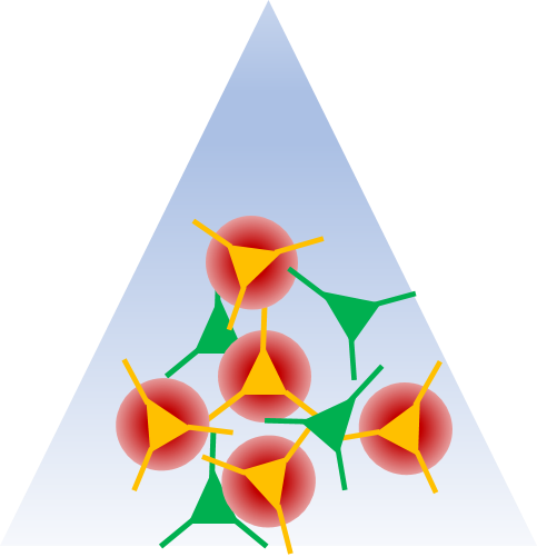
OptogeneticsOptogenetics is a great tool to modulate neural activity with millisecond time resolution. We use optogenetics to determine whether defined brain areas or cell types within a brain area are involved in normal arousal/sleep circuits or if alterations in these circuits can contribute to conditions such as cataplexy and REM sleep behaviour disorder.
|
|
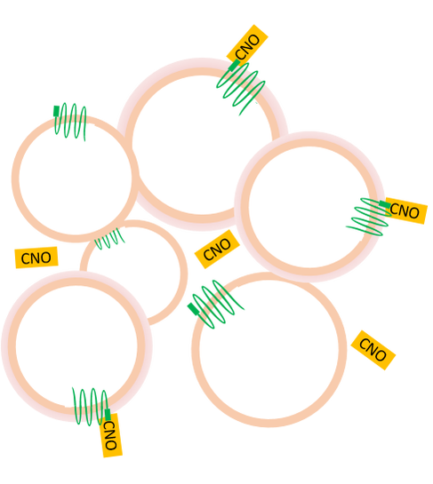
DREADDsDREADDs = Designer Receptors Exclusively Activated by Designer Drugs
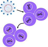
DREADDs also allow us to modulate the activity of brain regions or specific neurons. However, DREADDS allows us to do this over longer periods of time (e.g., hours-days). We use DREADDS to determine whether long-term suppression or activation of brain regions or cell types alters sleep/wake patterns or cycles. Viral VectorsViral vectors are a tool that allow us to introduce specific genes into defined brain regions. For example, for optogenetics, we can use a viral vector to introduce the gene for the light-sensitive opsin known as chanelrhodopsin. The cells influenced by the viral vector will then express this opsin which allows us to then turn these cells on by shining a light on them. In our lab we use viral vectors
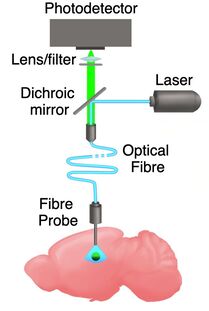
Fibre PhotometryFibre photometry is a method use to record the activity of specific cell type in any given brain region. It involves using light to stimulate and record the fluorescence of genetically encoded calcium indicators (GECIs). The fluorescence detected directly correlates with the cellular calcium signalling of neurons when they become active. Fibre photometry can also be used with optogenetics to temporally control the excitation or inhibition of selected population of neurons during the sleep/wake cycle.
|
|
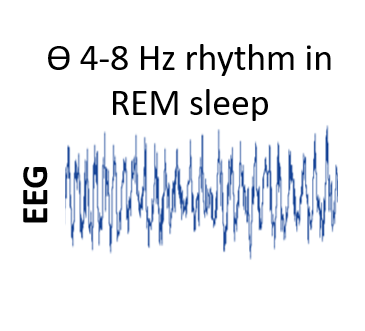
Electroencephalography (EEG)EEG recordings have been used in science since the 1950s and are still one of the best tools to distinguish different arousal states. We primarily use EEG recordings to determine what arousal state an animal is in (e.g., awake, REM sleep, non-REM sleep). We also record local field potential activity to determine whether specific features of the activity in a brain region correlate with an animal's arousal state.
|
|
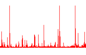
Electromyography (EMG)EMG refers to the technique that allows us to monitor and record the electrical activity generated by skeletal muscle. We typically record EMG activity in cheek and neck muscles to help determine what arousal state an animal is in.
|
|
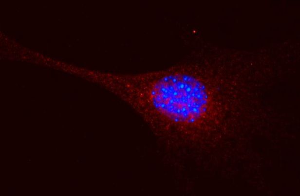
Cell CultureWith cell culture techniques you can grow different cells outside of a live body. We are interested in determining whether cells cultured outside the brain can be transplanted into a brain and restore normal function in individuals that lack these cells.
|
|
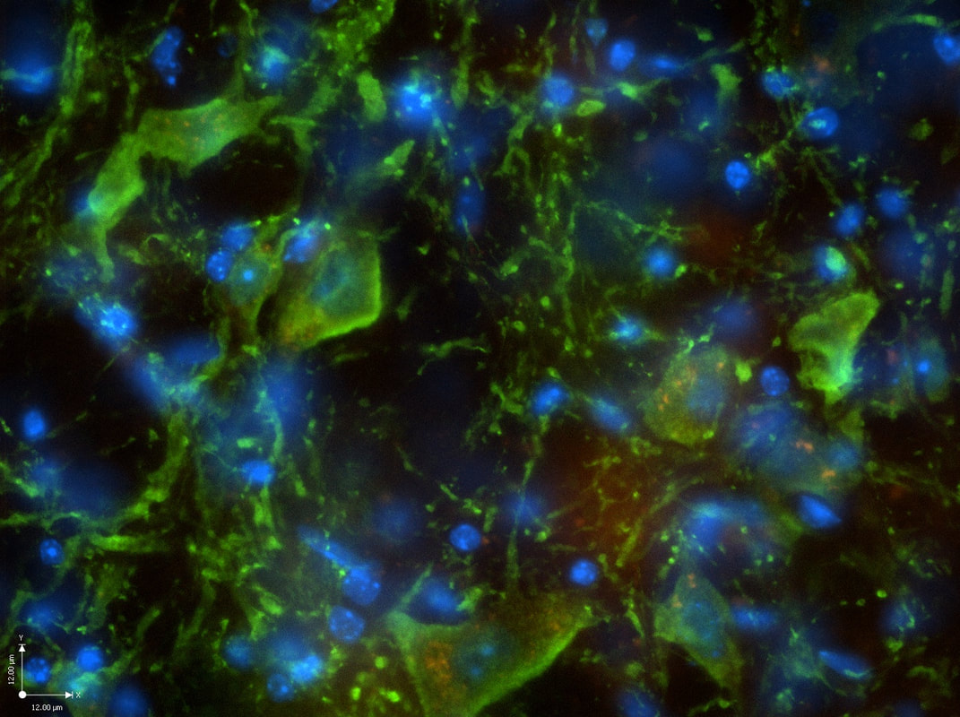
Molecular TechniquesWe use a variety of molecular biology techniques to detect the presence of various proteins, mRNA, or genes. To do this, we use techniques such as immunohistochemistry, western blotting, in situ hybridization, and polymerase chain reaction (PCR).
|